• 1. APRIL 2018
    Projection length stimulated by oxytocin is modulated by the inhibition of calcium signaling in U-87MG cells

    Projection length stimulated by oxytocin is modulated by the inhibition of calcium signaling in U-87MG cells

    AUTORI: Zatkova, M., Z. Bacova, F. Puerta, Z. Lestanova, M. Alanazi, A. Kiss, A. Reichova, A.M. Castejon, D. Ostatnikova, and J. Bakos

    ABSTRACT: Neuropeptide oxytocin contributes to the regulation of glial cell morphology. The precise mechanisms, however, are not yet fully understood. In the present study, we have investigated whether an oxytocin-induced increase of intracellular calcium is required for cell extension in astrocyte-like U-87MG cells. Oxytocin (1 µM) significantly increased the length of the cell projections measured by the green-fluorescent protein labeled microtubule-associated protein after 48 h. The knockdown of oxytocin receptors (OXTR) in U-87MG cells prevented the elongation of the projections. Incubation of U-87MG cells in the presence of oxytocin, resulted in a significant increase of intracellular calcium, specifically blocked by the OXTR antagonist L-371,257. Both quercetin, which is a phosphoinositide 3-kinase inhibitor, and the phospholipase C inhibitor U-73122 reduced oxytocin-induced elevation of intracellular calcium concentration. Conversely, neither diltiazem, an L-type voltage-gated calcium channel blocker nor tetracaine, which is a blocker of the ryanodine receptors, showed an effect on intracellular calcium levels. Treatment of cells with quercetin, U-73122 and the voltage-gated calcium channel blockers cilnidipine, ω-agatoxin and mibefradil prevented the elongation of projections stimulated by oxytocin. Oxytocin treatment resulted in a significant increase in gene and protein expression of the scaffolding protein SHANK3. Our results clearly show that activation of OXTRs contributes to the elongation of cell projections in astrocyte-like U-87MG cells and that this effect is mediated by an extracellular calcium influx accompanied by an increase in scaffolding proteins expression.

    J Neural Transm (Vienna), 2018. 125(12): p. 1847-1856.

    https://pubmed.ncbi.nlm.nih.gov/30269165/

    • 1. FEBRUARY 2018
    Etiopatogenéza autizmu – súčasný stav poznania (Etiopathogenesis of autism – current state of knowledge)

    Etiopatogenéza autizmu – súčasný stav poznania (Etiopathogenesis of autism – current state of knowledge)

    AUTORI: Važan, R., Belica, I., Ostatníková, D.

    ABSTRACT: Autism is a set of phenotypically heterogeneous neurodevelopmental conditions characterized by core symptoms in social communication, interactions, interests and repetitive stereotyped behaviour. Therefore, the term – autistic spectrum disorders, is preferred today. Due to the progress of autism in recent decades, a wide range of studies have been done to identify the etiology. Autism is very probably caused by multiple etiological factors affecting central nervous system development in critical periods of early life. High heritability suggests that genes play key role in autism etiology. A number of candidate genes, that control brain development, production of neurotransmitters and neuromodulators, have been identified. Gamma-aminobutyric acid, glutamate, dopamine, noradrenaline, serotonin, acetylcholine are in the centre of research interest as well as the hormones – testosterone, melatonin and oxytocin. Recently the interactions between susceptible genes and environmental factors have been proposed as the major
    mechanism of autism etiopathogenesis. This idea is supported by huge phenotypic diversity of individuals with autistic spectrum disorders.

    Lekársky obzor. – Roč. 67, č. 7-8 (2018), s. 234-238. – ISSN (print) 0457-4214

    Lekársky obzor – HERBA – Lekársky obzor 7-8/2018

    • 1. JUNE 2017
    Vitamin D, neurosteroids and autism

    Vitamin D, neurosteroids and autism

    AUTORI: Macova, L., M. Bicikova, D. Ostatnikova, M. Hill, and L. Starka

    ABSTRACT: Vitamin D had been for a long time investigated for its effects on
    bone metabolism. Recently has been observed that the incidence of some neurodevelopmental disorders (including autism) increases hand in hand with vitamin D deficiency. Indeed, vitamin D was reported to modulate the biosynthesis of neurotransmitters and neurotrophic factors; moreover, its receptor was found in the central nervous system. Vitamin D deficiency was therefore assessed as a risk factor for autism, however the biological mechanism has not yet been revealed. In our review we focused on potential connections among vitamin D, steroids and autism. Potential mechanisms of vitamin D action are also discussed.

    Physiol Res, 2017. 66(Supplementum 3): p. S333-S340.

    http://www.biomed.cas.cz/physiolres/pdf/66/66_S333.pdf

    • 1. JUNE 2017
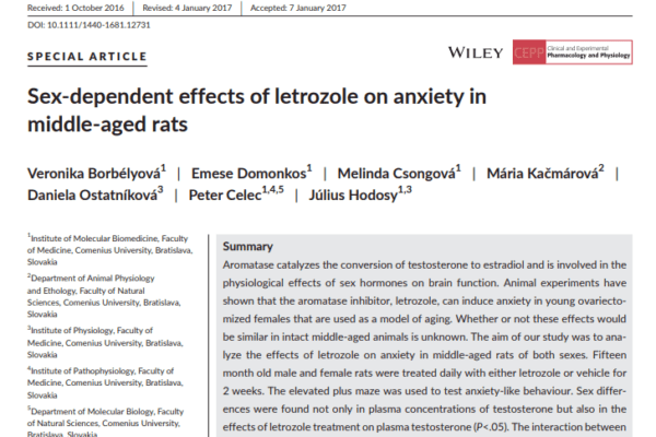
    Sex-dependent effects of letrozole on anxiety in middle-aged rats

    Sex-dependent effects of letrozole on anxiety in middle-aged rats

    AUTORI: Borbelyova, V., E. Renczes Domonkos, M. Csongova, M. Kacmarova, D. Ostatnikova, P. Celec, and J. Hodosy

    ABSTRACT: Aromatase catalyzes the conversion of testosterone to estradiol and is involved in the physiological effects of sex hormones on brain function. Animal experiments have shown that the aromatase inhibitor, letrozole, can induce anxiety in young ovariectomized females that are used as a model of aging. Whether or not these effects would be similar in intact middle-aged animals is unknown. The aim of our study was to analyze the effects of letrozole on anxiety in middle-aged rats of both sexes. Fifteen month old male and female rats were treated daily with either letrozole or vehicle for 2 weeks. The elevated plus maze was used to test anxiety-like behaviour. Sex differences were found not only in plasma concentrations of testosterone but also in the effects of letrozole treatment on plasma testosterone (P<.05). The interaction between sex and treatment was also proven in locomotor activity (P<.05) and time spent in the open arms of the elevated plus maze (P<.05). Letrozole-treated male rats spent 95% less time in the open arms of the elevated plus maze than the control rats did (P<.05) suggesting an anxiogenic effect of aromatase inhibition. This difference was not found between letrozole-treated and vehicle-treated females. In contrast to previous experiments on young animals, letrozole seems to induce anxiety in male but not in female middle-aged rats. This sex-specific effect might be related to sex differences of oestrogen and androgen signalling in aging brains. These results should be taken into account in clinical applications of letrozole, especially in men.

    Clin Exp Pharmacol Physiol, 2017. 44 Suppl 1: p. 93-98.

    https://onlinelibrary.wiley.com/doi/full/10.1111/1440-1681.12731